A Guide to Clinical Differential Diagnosis of Oral Mucosal Lesions
Course Number: 110
Course Contents
White Lesions Due to Subepithelial Change
White lesions due to subepithelial change have normal overlying epithelium, but changes in the connective tissue partially mask blood vessels and cause the area to appear white, yellow or tan. These lesions have a smooth translucent surface, do not rub off, and are not painful.
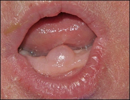
Gingivial cyst of the newborn* is also known as dental lamina cyst of the newborn. This is an epithelial inclusion cyst found on the attached alveolar mucosa of infants. It presents as an asymptomatic white thickened surface lesion. Similar cysts occur on the hard palate. No treatment is necessary as the lesions resolve spontaneously within several weeks after birth.
Gingivial cyst of newborn
Gingivial cyst of newborn
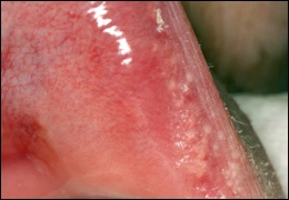
Fordyce granules* appear as flat or slightly elevated, yellow clusters, most commonly located on the buccal mucosa and lip. They represent sebaceous glands. Fordyce granules are harmless and require no treatment.
Fordyce granules
Fordyce granules
Scarring of the oral mucosa can appear as white surface lesions with a smooth surface. They are non-painful and do not rub off. Diagnosis is made by history of trauma or surgery to the area. No treatment is necessary.
To view the Decision Tree for Oral Mucosal Lesions, click on one of the options shown.
To view the Decision Tree for Oral Mucosal Lesions, click on one of the options shown.


 View Interactive
 View as PDF
 View as GIF